|
|
|
|
 -
- 
Conference
on Mathematics of Medical Imaging
June 20-24, 2011
hosted by the Fields Institute
held at the University of Toronto |
Organizing
Committee:
Adrian Nachman , University of Toronto
Dhavide Aruliah, University of Ontario Institute of Technology
Hongmei Zhu, York University
|
| |
Research Posters
- Alex
Martinez
Simulation of Magnetic Resonance Imaging using Oscillatory Quadrature
Methods
- Nargol Rezvani
A Polyenergetic Iterative Reconstruction Framework for X-Ray Computerized
Tomography
- Mihaela Pop
Experimental framework to parameterize 3D MR image-based computer
models of electrophysiology in heterogeneous infarcted porcine
hearts
- Dinora Morales
Spatial clustering analysis of functional magnetic resonance
imaging data
- Lili Guadarrama
Bustos
Transient Wave Imaging
-
Golafsoun Ameri, Eric
Strohm, Carl Kumaradas, Victor Yang
Synthetic Aperture Imaging in Acoustic Microscopy
-
N.
Tabatabaei, A. Mandelis,
Thermophotonic Radar Imaging of Turbid Media
-
Bahman Lashkari,
and Andreas Mandelis,
Photoacoustic wave generation and signal-to-noise ratio modeling
-
C. Liu, W. Gaetz, T.P.L.
Roberts, and H. Zhu,
Assessing the Functional Significance of MEG Motor Cortex Gamma
Oscillations Using Time-frequency Analysis
- Evgeniy Lebed, Mei Young,
Yifan Jian, Paul J. Mackenzie, Marinko V. Sarunic, Mirza Faisal
Beg
Real time Compressive Sampling based FDOCT image acquisition
and registration
Simulation
of Magnetic Resonance Imaging using Oscillatory Quadratures
Methods
by
Alex Martinez
University of Toronto
Coauthors: Luca Antiga (Orobix Srl and Mario Negri Institute),
David Steinman (University of Toronto)
Magnetic resonance imaging (MRI) has
become one of the leading modalities for non-invasive anatomical
imaging. However, there are many independent parameters that
control an MRI scan and many physical phenomena that affect
the quality and accuracy of the acquired image. Studying the
causes and effects of these phenomena is difficult, because
MRI facility availability is scarce and operating time is costly.
Computational simulation is MRI has become an attractive alternative,
but can suffer from extensive simulation times. Moreover simulations
are usually based on structured, Cartesian grids, which must
be very dense in order to adequately resolve anatomically realistic
objects.
An alternative approach has been suggested
in which the MRI signal equation, which represents the volumetric
integration of a magnetized object modulated by a sinusoidally
varying field, can be solved exactly over objects defined by
an unstructured grid of linear tetrahedral elements [1]. If
an object can be segmented into regions over which each a constant
magnetization can be assumed, the signal for these regions can
be converted, via the divergence theorem, into the result of
a surface integration over linear triangles [2]. In either case,
however, the number of simplexes, and hence the CPU time, required
to resolve the curved boundaries of realistic objects, can be
prohibitive.
The present work focuses on the use of
quadratic triangulations, which have been shown to offer significant
reductions in the number of simplexes required to discretize
complex objects [3], but which require numerical rather than
exact integration of the signal equation. Due to the oscillatory
terms in the signal equation, conventional Gaussian quadratures
can be costly, as the number of points needed in each dimension
is proportional to the maximum spatial frequency in the simulation.
Instead, we consider here the novel use of highly oscillatory
quadratures, for which the number of integration points decreases
with increasing frequency. Specifically, in the numerical steepest
descent (NSD) approach [4], the path between the integration
limits is deformed using the method of stationary phase, but
instead of trying to find an asymptotic estimate of the integral
afterwards, the new integral is evaluated using Gaussian quadrature.
This method can then be applied recursively for integrals of
n dimensions.
For a given number of integration points
the NSD approach can be expected to yield lower errors compared
to Gaussian quadrature. However, preliminary estimates suggest
that each NSD quadrature point require 3-4 times the number
of operations compared Gaussian quadrature. Moreover, NSD requires
special handling for some combinations of simplex and spatial
frequency orientations [4]. We intend to demonstrate whether
the perceived benefits of oscillatory vs. conventional quadratures
for simulating MRI are outweighed by these extra computational
costs.
References:
Truscott KJ and Buonocore MH. Simulation
of tagged MR images with linear tetrahedral solid elements.
J Magn Reson Imaging 2001;14:336-340.
Antiga L and Steinman DA. Efficient MRI
simulation via integration of the signal equation over triangulated
surfaces. Proc Int Soc Magn Reson Med 2008;16:489.
Simedrea P, Antiga L, Steinman DA. FE-MRI:
Simulation of MRI using arbitrary finite elements. Proc Int
Soc Magn Reson Med 2006:14:2946.
Huybrechs D and Vandewalle S. The construction
of cubature rules for multivariate highly oscillatory integrals.
Math Comp 2007; 76:1955
Back to Top
A Polyenergetic
Iterative Reconstruction Framework for X-Ray Computerized Tomography
by
Nargol Rezvani
Department of Computer Science, University of Toronto
Coauthors: D. A. Aruliah, Kenneth R. Jackson
While most modern x-ray CT scanners rely
on the well-known filtered back-projection (FBP) algorithm,
the corresponding reconstructions can be corrupted by beam-hardening
artifacts. These artifacts arise from the unrealistic physical
assumption of monoenergetic x-ray beams. To compensate, we discretize
an alternative model directly that accounts for differential
absorption of polyenergetic x-ray photons. We present numerical
reconstructions based on the associated nonlinear discrete formulation
incorporating various iterative optimization frameworks.
Back to Top
Experimental
framework to parameterize 3D MR image-based computer models
of electrophysiology in heterogeneous infarcted porcine hearts
by
Mihaela Pop
Sunnybrook Research Institute, Toronto
Coauthors: Maxime Sermesant (INRIA, France) Tommaso Mansi (Siemens
Corporate Research, Princeton, USA) Sudip Ghate (Sunnybrook
Research Institute, Toronto) Jean-Marc Peyrat (Siemens Molecular
Imaging, Oxford, UK) Jen Berry (Sunnybrook Research Institute,
Toronto) Beiping Qiang (Sunnybrook Research Institute, Toronto)
Elliot McVeigh (Johns Hopkins University, USA) Eugene Crystal
(Sunnybrook Research Institute, Toronto) Graham Wright (Sunnybrook
Research Institute, Toronto)
Mathematical modelling, high-resolution
imaging and electrophysiology experiments are needed to better
understand how tissue heterogeneities contribute to the genesis
of arrhythmia in hearts with prior infarction (a major cause
of sudden cardiac death). The purpose of this work was to globally
parameterize a 3D magnetic resonance MR image-based computer
model of electrophysiology (EP) constructed using a pre-clinical
pig model of chronic infarct. The computer heart model was built
from high-resolution ex-vivo 3D MRI scans. Diffusion weighted
MRI was used to estimate myocardial anisotropy (i.e., fiber
directions) and heterogeneities (healthy zone, dense scar and
border zone, BZ). We used a simple mathematical model based
on reaction-diffusion equations, and calculated the propagation
of action potential (AP) after application of stimuli (with
location and timing replicating precisely the stimulation protocol
used in the experiment). Specifically, the mathematical parameters
were globally fit by zone (i.e., the three zones derived from
heterogeneous MRI maps); this step was performed using characteristics
of AP waves measured ex-vivo (using 2D optical fluorescence
imaging). Then, these fitted parameters were further used as
input to the 3D computer model to replicate in-vivo EP studies,
under pacing or arrhythmia induction. Our results showed a better
agreement between experiments and simulations, when these customized
parameters were used instead of literature values. Future work
will focus on constructing the model from in-vivo MR images
and translating the model into clinical applications.
Back to Top
Spatial clustering
analysis of functional magnetic resonance imaging data
by
Dinora Morales
Universidad Politécnica de Madrid
Coauthors: Concha Bielza, Pedro Larrañaga
Functional magnetic resonance imaging
(fMRI) allows the brain function detection by measuring hemodynamic
changes related to neuronal activity given stimulus or task.
The central problem in the analysis of fMRI is the reliable
brain activated detection. One way is to compute a statistical
map and the spatial dependence among voxels are making during
inference form it. Clustering techniques have been applied to
statistical map based on extent of activation cluster after
intensity thresholding or taking into account contextual information
clustering. In this paper we focus on the spatial information
of fMRI to detect the brain activity taking into the spatial
contiguity constraints using the neighbourhood expectation maximization
algorithm with four and eight neighbourhood configurations.
The neighbourhood expectation minimization algorithm was applied
to Alzheimer's disease fMRI study.
Back to Top
Transient
Wave Imaging
by
Lili Guadarrama Bustos
Laboratoire de Mathematiques, Universite Paris-Sud 11. France
We study Elasticity imaging by the use
of the acoustic radiation force of an ultrasonic focused beam
to remotely generate mechanical vibrations in organs.We
provide a solid mathematical foundation for this transient technique
and design accurate methods for anomaly detection using transient
measurements.
We consider transient imaging in a non-dissipative
medium. We develop anomaly reconstruction procedures that are
based on rigorously established inner and outer time-domain
asymptotic expansions of the perturbations in the transient
measurements that are due to the presence of the anomaly.
Using the outer asymptotic expansion,
we design a time-reversal, Kirchhoff-, MUSIC- imaging technique
for locating the anomaly. Based on such expansions, we propose
an optimization problem for recovering geometric properties
as well as the physical parameters of the anomaly.
In the case of limited-view transient
measurements, we construct Kirchhoff- and MUSIC- algorithms
for imaging small anomalies. Our approach is based on averaging
of the limited-view data, using weights constructed by the geometrical
control method; It is quite robust with respect to perturbations
of the non-accessible part of the boundary. Our main finding
is that if one can construct accurately the geometric control
then one can perform imaging with the same resolution using
partial data as using complete data.
Back to Top
Synthetic Aperture
Imaging in Acoustic Microscopy
Golafsoun Ameri, Eric Strohm, Carl Kumaradas, Victor Yang
Acoustic microscopy (AM) provides micro-meter resolution using
a highly focused single-element transducer. A drawback in AM
is a relatively small depth of filed, resulting in poor resolution
outside the focus. Synthetic aperture (SA) image reconstruction
techniques can be used to improve the image resolution throughout
the field of view. SA mathematically synthesizes the effect
of an array transducer and produces dynamic focusing and depth-independent
resolution. SA reconstructions in both time domain (TD) and
frequency domain (FD) were implemented and tested using simulated
and experimental radio-frequency data from an acoustic microscope
at 400 MHz. Lateral resolutions of the SA reconstructed images
were better than those of conventional B-mode images. While
both TD and FD algorithms improved the resolution, the FD algorithm
had better resolution. In conclusion, FD-SA improves resolution
in AM outside the focal region, at the expense of real-time
imaging.
Back to main index
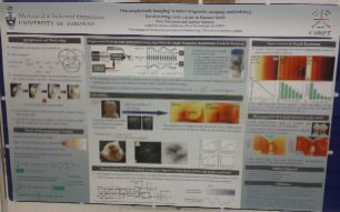
Thermophotonic
Radar Imaging of Turbid Media
by N. Tabatabaei*, A. Mandelis**
*Center for Advanced Diffusion-Wave Technologies (CADIFT), MIE
Dept., University of Toronto, Toronto (Ontario), Canada M5S 3G8,
nimat@mie.utoronto.ca
** Center for Advanced Diffusion-Wave Technologies (CADIFT), MIE
Dept., University of Toronto, Toronto (Ontario), Canada M5S 3G8,
mandelis@mie.utoronto.ca
Lock-in thermography is an active thermographic
method that incorporates quadrature demodulation to retrieve the
amplitude and phase of the thermal-waves generated inside the
sample either optically, acoustically or mechanically. The role
of subsurface defects, in this case, is then to shift the thermal-wave
centroid and therefore produce a contrast, both in amplitude and
phase images, with respect to the intact areas. The significant
difference of biological samples (turbid media) is that due to
their translucency the infrared radiation emanating from them
is governed by a coupled diffused-photon-density and thermal-wave
field ("thermophotonics"), as opposed to purely thermal-wave
field in opaque materials:
Optical field: ;
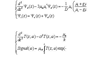
Thermal field:
The case of biological samples is a challenging case as these
samples are usually translucent and do not effectively absorb
the applied optical excitation. Even if they do, medical safety
codes prevent researchers from applying high power excitation
to these samples. As a result, the photothermal signals obtained
from biological samples are generally poor in terms of signal-to-noise
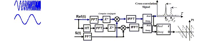
ratio (SNR). The intension of this poster presentation is to investigate
the use of matched-filter Radar processing in the thermophotonic
imaging of turbid media That is, the optical excitation is performed
in a linear frequency modulated (chirped) or binary phase-coded
manner and the infrared response from the sample is matched-filtered
to the applied excitation according to the algorithm below:
One immediate outcome of such methodology
is the ability to form depth-selective ( =constant) images rather
than lock-in thermography's depth integrated images as well as
maintaining higher SNR and axial resolution. The figure below
compares the phase images obtained from a classic step-wedge sample
inside a scattering phantom. The results clearly show the enhanced
axial resolution of Radar imaging compared to that of the conventional
lock-in imaging. 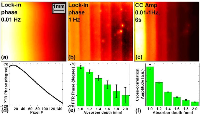
This poster presentation provides the analytical
solution to the thermophotonic Radar problem of an absorber in
a turbid medium and verifies the capabilities of the proposed
methodology through detection of early dental caries in human
teeth.
Back to Top
Photoacoustic
wave generation and signal-to-noise ratio modeling
Bahman Lashkari, and Andreas Mandelis
Center for Advanced Diffusion-Wave Technologies (CADIFT), Department
of Mechanical and Industrial Engineering, University of Toronto,
Toronto, M5S 3G8, Canada
The generation of photoacoustic (PA) transients was modeled by
employing a two dimensional axially symmetric solution in the
frequency-domain. The frequency-domain solution facilitates the
incorporation of the transducer dynamic and acoustic attenuation
effects. In addition, the two- layer model automatically introduces
the implementation of an arbitrary acoustic boundary condition.
It has been shown that this solution asymptotically approaches
the one-dimensional solution under specific conditions for beam
spotsize and/or absorber size and minimum excitation frequency.
The model has been used for both pulsed and continuous wave (CW)
PA to predict the maximum signal and signal-to-noise ratio (SNR).
In the CW PA, many parameters can be manipulated to increase the
detected signal. The most important parameter is the frequency
bandwidth of the excitation energy. The developed model predicts
the optimum parameters to maximize the SNR. This analysis also
provides a relative formulation depending on utilized parameters
for the study of the performance of both modalities. This relative
performance formulation demonstrates that by judicious selection
of the chirped FD PA parameters, this method is capable of competing
with the pulsed PA counterpart to generate superior SNR and resolution.
The theoretical predictions were compared with experimental results
achieved for both modalities using a dual-mode PA system.
Back to Top
Assessing the Functional Significance
of MEG Motor Cortex Gamma Oscillations Using Time-frequency Analysis
C. Liu1, W. Gaetz2, T.P.L. Roberts2, and H. Zhu1
1. Department of Mathematics and Statistics, York University,
Toronto, ON, Canada
2. Lurie Family Foundation MEG Imaging Center, Department of Radiology,
Children's Hospital of Philadelphia, Philadelphia, PA, United
States
Gamma-band responses (40-90 Hz) are thought to represent a key
neural signature of information processing in the human brain.
Motor gamma band responses have also been observed for brief periods
typically observed around movement onset, yet the functional significance
of these responses remains unclear. In this study, we investigate
the influence of task difficulty on the gamma-band motor cortex
activity using the multi-source interference task (MSIT), a task
designed in maximizing response interference. Due to huge variations
of dynamic structures of brain functional activity, we propose
an adaptive time-frequency analysis tool whose time-frequency
resolution is adaptively adjusted to its analyzed signal; thus
more accurate description of local signal characteristics can
be obtained.
Fifteen right-handed subjects performed the MSIT. 80 control and
80 interference trials were recorded for each subject. Brain activity
was recorded continuously using a 275 channel whole-head magnetoencephalography
(MEG) (1200 samples/s). A differential minimum-variance beamformer
algorithm was applied to identify the location of gamma-band (60-90
Hz) activity at the contralateral primary motor cortex (MIc).
The proposed time-frequency analysis technique was applied to
single trial MEG data from peak gamma-band locations. Gamma-band
activity revealed in the time-frequency domain was compared for
control and interference trials, and then for fast and slow trials,
respectively.
Analysis results suggest that MIc gamma is significantly active
for responses requiring relatively more processing time (slow
vs. fast trials), and for tasks within the interference condition
(interference vs. control trials). Anatomical connections between
MI cortex and sub-thalamic nucleus (STN) are well known, and STN
is also known to exhibit activity in gamma band. Thus, the current
results may suggest enhanced MIc to STN communication with increasing
task demands such as with the MSIT task.
Operator Independent Transcranial
Doppler Ultrasound for Continuous Monitoring of Cerebral Vessels
(poster image)
Lee B., Kumaradas JC, Yang V, Ryerson University
Continuous monitoring of the blood vessels 3-14 days after subarachnoid
hemorrhage (SAH) from cerebral aneurysm rupture is imperative
to assess the presence of vasospasms. Transcranial Doppler Ultrasound
(TCD) can now be used for continuous monitoring of vasospasm.
However, the use of TCD suffers from operator dependence requiring
a skilled ultrasonographer to make doppler angle corrections.
The aim of the research is to minimize the need of dedicated ultrasonographers
for TCD monitoring of cerebral vasospasms. The 3D vascular structure
of a phantom was obtained using binary skeletonization from 3D
power Doppler images. The vascular structure was used in combination
with angle independent pulsed Doppler to reconstruct the temporal
blood velocity profiles at various parts of the vasculature. The
results indicate the operator independent monitoring of cerebral
vasospasm is possible.
Back to main index
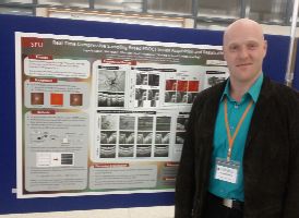
Real time Compressive Sampling based FDOCT
image acquisition and registration
Evgeniy Lebed, Mei Young, Yifan Jian, Paul J. Mackenzie,
Marinko V. Sarunic, Mirza Faisal Beg
Purpose Acquiring Fourier Domain Optical Coherence Tomography
(FDOCT) at high speed is becoming an important problem in ophthalmic
imaging. We present a medical imaging interpolation technique
called Compressive Sampling (CS) for rapid volumetric acquisition
of retina and Optic Nerve Head (ONH) in humans and in rodents.
Methods: The 3D volumes were acquired with a custom FDOCT system.
A reduction in the acquisition time was implemented by modification
of the scan pattern to acquire only a subset of the area (up to
only 25%) using randomly spaced horizontal and vertical B-scans.
Compressive sampling techniques were used to interpolate the missing
data with high fidelity for scan time reductions of up to 73%
on human ONH volumetric data.
Results: Reconstructions using the Compressive Sampling (CS) method
were performed on sparsely acquired human retinal images. We show
that it is possible to obtain several sparsely-acquired volumes
in the same time that it would take to acquire a fully-sampled
volume, and by means of non-rigid registration we obtain volumetric
images that are potentially more preferential than the fully-sampled
FDOCT images.
Conclusions: We demonstrated that Compressive Sampling can be
used to reconstruct 3D FDOCT images with minimal degradation in
quality. We showed that there is negligible effect on human retinal
layers and on clinically relevant morphometric measurements of
the human ONH. We also demonstrate that there is a significant
reduction in motion artifacts when we sparsely sample the volume.
The potential outcome of this work is a significant reduction
in FDOCT image acquisition time for clinical volumetric imaging
applications.
Back to main index
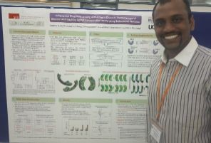
A Comprehensive Study of Differential
Diagnosis among Alzheimer's Disease, Frontotemporal Disease and
Healthy Aging
Pradeep Kumar Raamana , Mirza Faisal Beg
Purpose: Alzheimer's disease (AD) and Frontotemporal dementia
(FTD) are challenging to discriminate due to large overlap in
clinical symptoms and the cognitive domains impaired. The NINCDS-ADRDA
criteria for diagnosing probable AD have a sensitivity of 93%
but a specificity of only 23% in distinguishing it from FTD as
most patients with FTD also fulfilled NINCDS-ADRDA criteria for
AD. Since pharmacologic treatments differ for AD and FTD, misdiagnosed
patients will incur side effects for no benefit with important
negative consequences. We present a comprehensive study in discriminating
among Alzheimer's disease, Frontotemporal disease and Healthy
Aging (HA) using various biomarkers.
Methods: The different biomarkers we compare and contrast are
volumes, shape, and surface displacements of both hippocampi and
lateral ventricles. The volumes and shape features are computed
from the binary segmentations obtained via multi-atlas fusion
of the segmentations from a cohort of a 30 FTD patients, 34 Probable
AD patients and 14 age-matched controls.
Results: All the biomarkers are studied in a 3-class setting
(AD, FTD and HA) using a fixed classifier to obtain the diagnostic
value of these biomarkers in the context of differential diagnosis.
To date, such a comprehensive study in a 3-class setting hasn't
been published to the best of our knowledge. A highlight of this
study is evidence of high diagnostic value of the ventricular
degeneration, in shape and deformation, for the differential diagnosis
of FTD, AD and HA. The results present a valuable insight into
the discriminative power of different biomarkers studied here
and demonstrate the potential of ventricular degeneration as biomarker
in the differential diagnosis of FTD, AD and HA.
Back to main index
|
|